hand xray anatomy
Radiographic Anatomy of the Skeleton: Lumbar Spine -- Lateral View we have 9 Pictures about Radiographic Anatomy of the Skeleton: Lumbar Spine -- Lateral View like Image | Radiopaedia.org, Rheumatoid arthritis hands | Radiology Case | Radiopaedia.org and also Image | Radiopaedia.org. Here it is:
Radiographic Anatomy Of The Skeleton: Lumbar Spine -- Lateral View
 uwmsk.org
spine lumbar lateral anatomy ray labelled radiology diagram coccyx radiographic xray human skeleton bones uwmsk muscle foot normal nursing schools
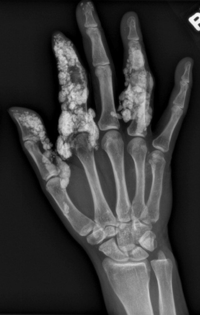
Scleroderma - Radiology At St. Vincent's University Hospital
 www.svuhradiology.ie
scleroderma radiology ray bone svuhradiology ie st radiograph
4th Metacarpal Fracture | Image | Radiopaedia.org
 radiopaedia.org
metacarpal fracture 4th lateral views radiopaedia
Normal Chest X-ray: Anatomy Tutorial | Kenhub
:background_color(FFFFFF):format(jpeg)/images/article/en/normal-chest-x-ray/eIIXNhFk2r9VNLRTkmISA_Screenshot_2019-02-21_at_10.07.34.png) www.kenhub.com
www.kenhub.com
ray chest normal anatomy kenhub
Boxer Fracture | Image | Radiopaedia.org
 radiopaedia.org
fracture boxer break knuckle boxers pinky finger metacarpal fractures 5th endocrine radiopaedia spring christmas version
Rheumatoid Arthritis Hands | Radiology Case | Radiopaedia.org
 radiopaedia.org
arthritis rheumatoid hands psoriatic ray hand radiopaedia ra radiology rash inflammatory psoriasis case
Carpal Bones - Wikipedia
 en.wikipedia.org
bones carpal xray hand carpals short human between difference bone anatomy carpus wiki trapezium wrist lunate scaphoid name hamate letter
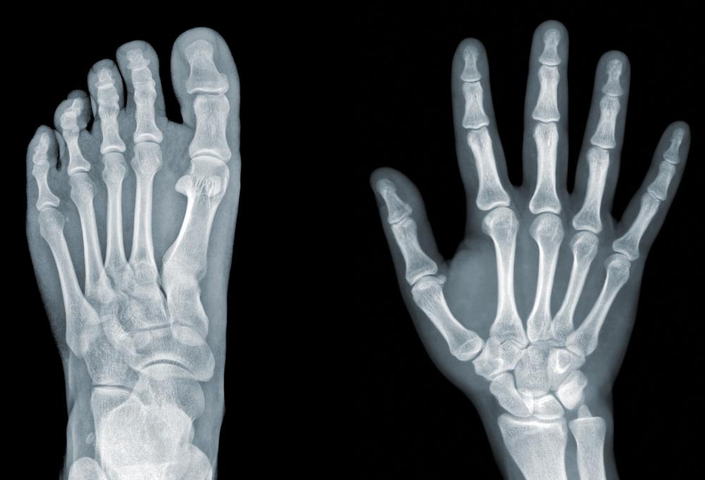
What Is The Difference Between A CT Scan And An X-Ray?
 www.wisegeek.com
ray rays ct scan does technician lead difference body equipment parts bones machines radiation community radiologist use internal detailed donegal
Image | Radiopaedia.org
 radiopaedia.org
ray radiopaedia
Scleroderma radiology ray bone svuhradiology ie st radiograph. Fracture boxer break knuckle boxers pinky finger metacarpal fractures 5th endocrine radiopaedia spring christmas version. Rheumatoid arthritis hands