lateral cxr anatomy
Malpositioned nasogastric tube - Radiology at St. Vincent's University we have 9 Images about Malpositioned nasogastric tube - Radiology at St. Vincent's University like How to Interpret a Chest X-Ray (Lesson 2 - A Systematic Method and, How to Interpret a Chest X-Ray (Lesson 5 - Cardiac Silhouette and and also Congenital diaphragmatic hernia | Image | Radiopaedia.org. Read more:
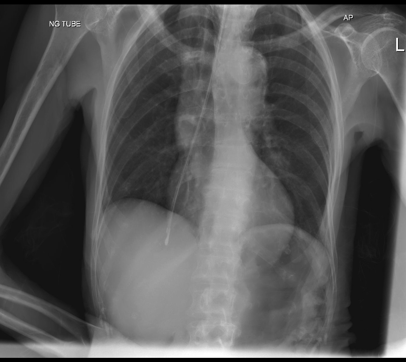
Malpositioned Nasogastric Tube - Radiology At St. Vincent's University
 www.svuhradiology.ie
tube nasogastric malpositioned radiology svuhradiology ie
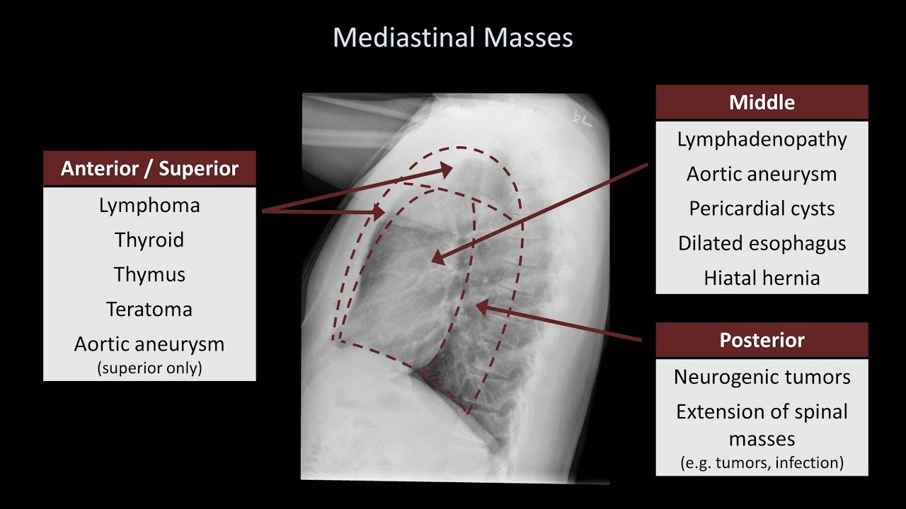
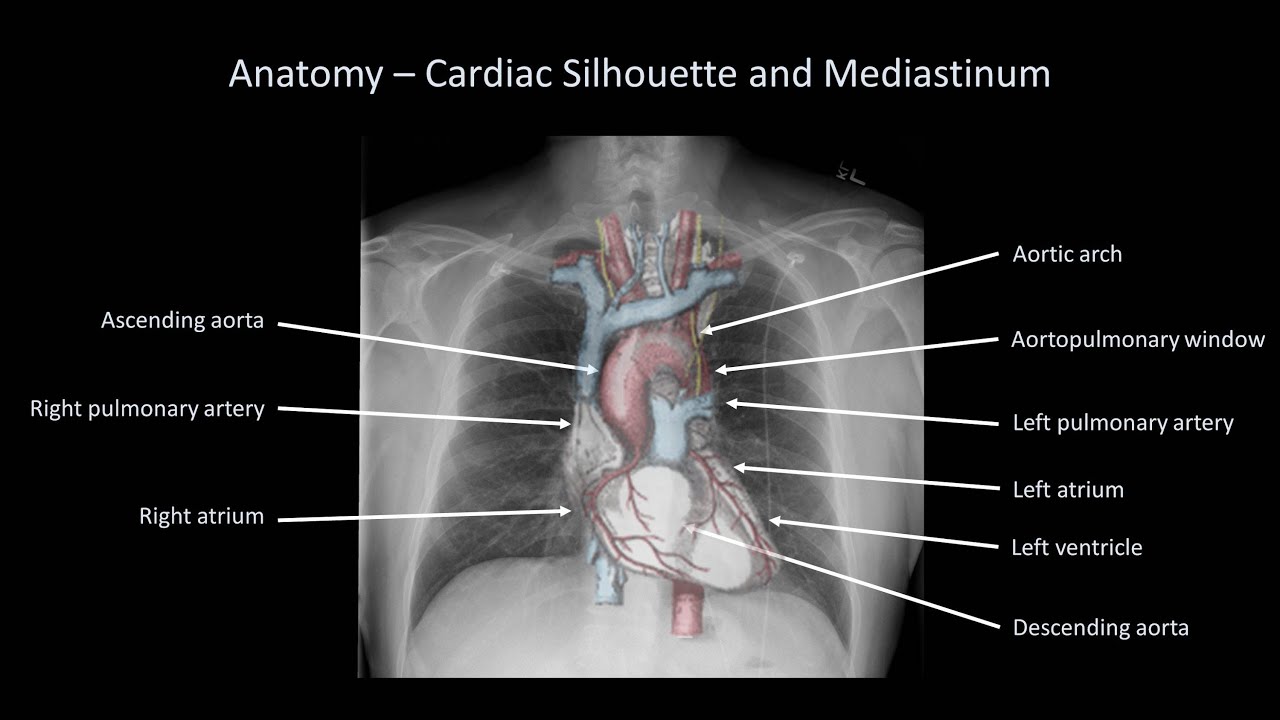
How To Interpret A Chest X-Ray (Lesson 5 - Cardiac Silhouette And
 www.youtube.com
chest ray cardiac silhouette mediastinum interpret lesson
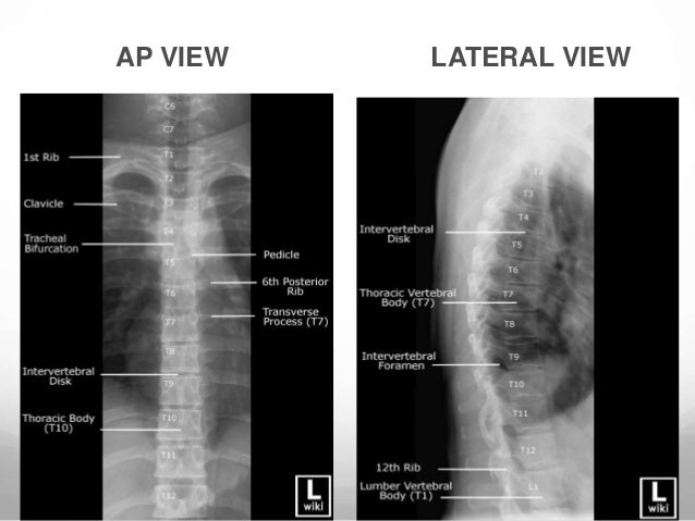
RADIOGRAPHY OF CHEST AND SPINE
 www.slideshare.net
radiography t11 distal lumbar intervertebral sacrum spinous vertebral
Congenital Diaphragmatic Hernia | Image | Radiopaedia.org
 radiopaedia.org
hernia diaphragmatic congenital lateral radiopaedia radiology remaining frontal case
Differentiate Left And Right Hemidiaphragms On Chest X-ray Lateral View
 www.youtube.com
lateral chest ray right left differentiate
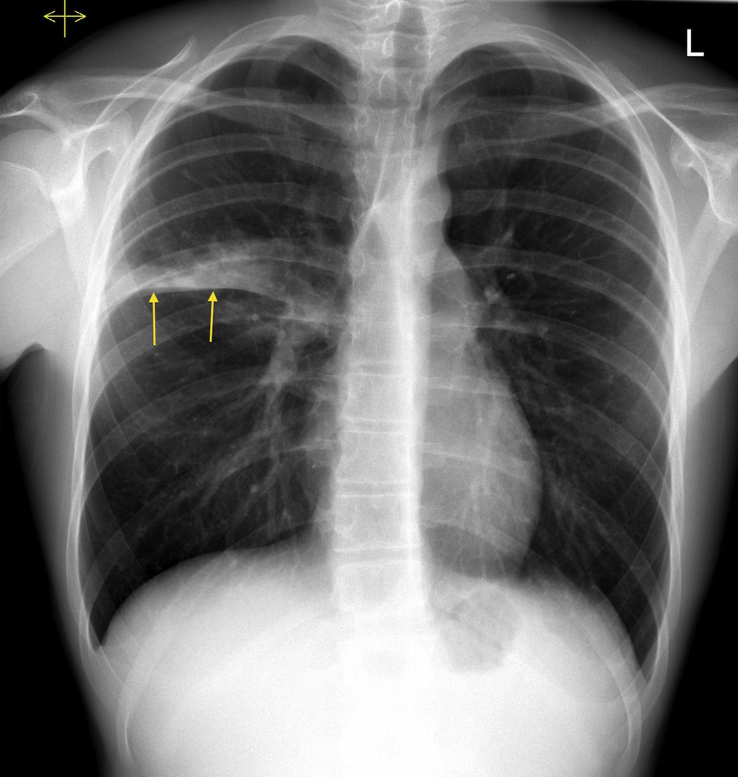
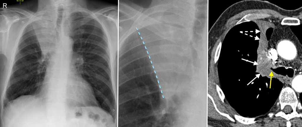
Right Upper Lobe Consolidation – CXR - Radiology At St. Vincent's
 www.svuhradiology.ie
lobe consolidation cxr upper right lung fissure horizontal radiology zone patient inferior margin
Left Lower Lobe Pneumonia - Lateral CXR - Radiology At St. Vincent's
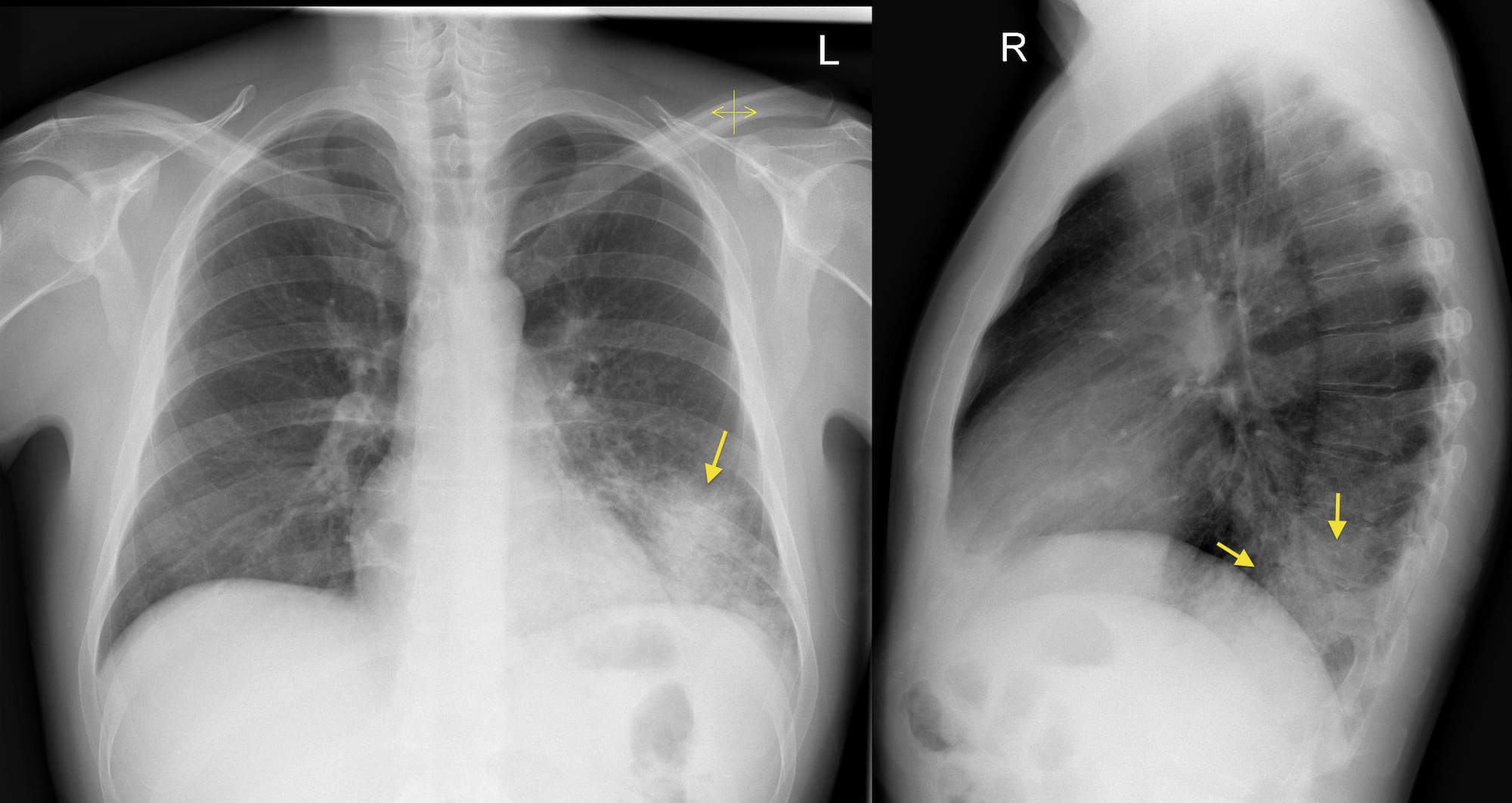 www.svuhradiology.ie
pneumonia lateral lobe cxr lower left right chest ray lobar pa views side film stepwards spine interpreting radiology
Right Upper Lobe Collapse - CXR/CT - Radiology At St. Vincent's
 www.svuhradiology.ie
lobe collapse upper right cxr ct radiology svuhradiology ie
How To Interpret A Chest X-Ray (Lesson 2 - A Systematic Method And
 www.youtube.com
chest ray anatomy interpretation radiology imaging easy method radiologic left healthy visit
Left lower lobe pneumonia. Malpositioned nasogastric tube. Lobe consolidation cxr upper right lung fissure horizontal radiology zone patient inferior margin