leg venous anatomy
(PDF) Management of Patients With Venous Leg Ulcers: Challenges and we have 8 Images about (PDF) Management of Patients With Venous Leg Ulcers: Challenges and like Veins of the upper limb: Anatomy | Kenhub, DVT ultrasound protocol. Step by step guide to ruling out a DVT and also Ultrasound Leadership Academy: Lower Extremity DVT — EM Curious. Here you go:
(PDF) Management Of Patients With Venous Leg Ulcers: Challenges And
 www.researchgate.net
leg management ulcers patients venous challenges practice current
Asian American Chinese Male Surface Anatomy Of The Leg Ankle And Foot
 joelgordon.photoshelter.com
DVT Ultrasound Protocol. Step By Step Guide To Ruling Out A DVT
 www.angiologist.com
ultrasound venous dvt veins vein calf peroneal protocol tibial posterior rule fill duplex angiologist
Vascular Anatomy Of Groin Medical Illustration Medivisuals
 medivisuals1.com
anatomy groin neurovascular vascular femoral arteries illustration pelvis medical veins vein medivisuals1 nerve artery system circulatory saphenous superficial
Upper Extremity Venous Doppler – Sonographic Tendencies | Vascular
 www.pinterest.com
extremity doppler venous ultrasound vascular vein sonographictendencies sonography tendencies sonographic thrombosis jugular
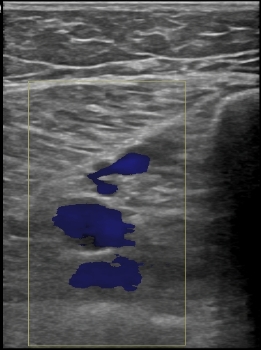
Ultrasound Leadership Academy: Lower Extremity DVT — EM Curious
 www.emcurious.com
dvt ultrasound study bedside lower extremity anatomy blood leadership accuracy academy am fullsize sinaiem shot screen
Veins Of The Upper Limb: Anatomy | Kenhub
:background_color(FFFFFF):format(jpeg)/images/article/en/veins-of-the-upper-limb/kBrClIBlNSnH1mqv529Vg_OMapCc29PWZK3PoeVttkw_qT4qkk7cT5_Vena_brachialis_2.png) www.kenhub.com
www.kenhub.com
veins upper limb vein brachial kenhub anatomy rhomboid calvaria arm ventral muscles deep venous superficial vena brachialis function library bones
Upper Extremity Venous Exam Technique And Interpretation - YouTube
 www.youtube.com
upper extremity venous interpretation exam
Dvt ultrasound protocol. step by step guide to ruling out a dvt. Upper extremity venous exam technique and interpretation. Vascular anatomy of groin medical illustration medivisuals