medial lateral anatomy
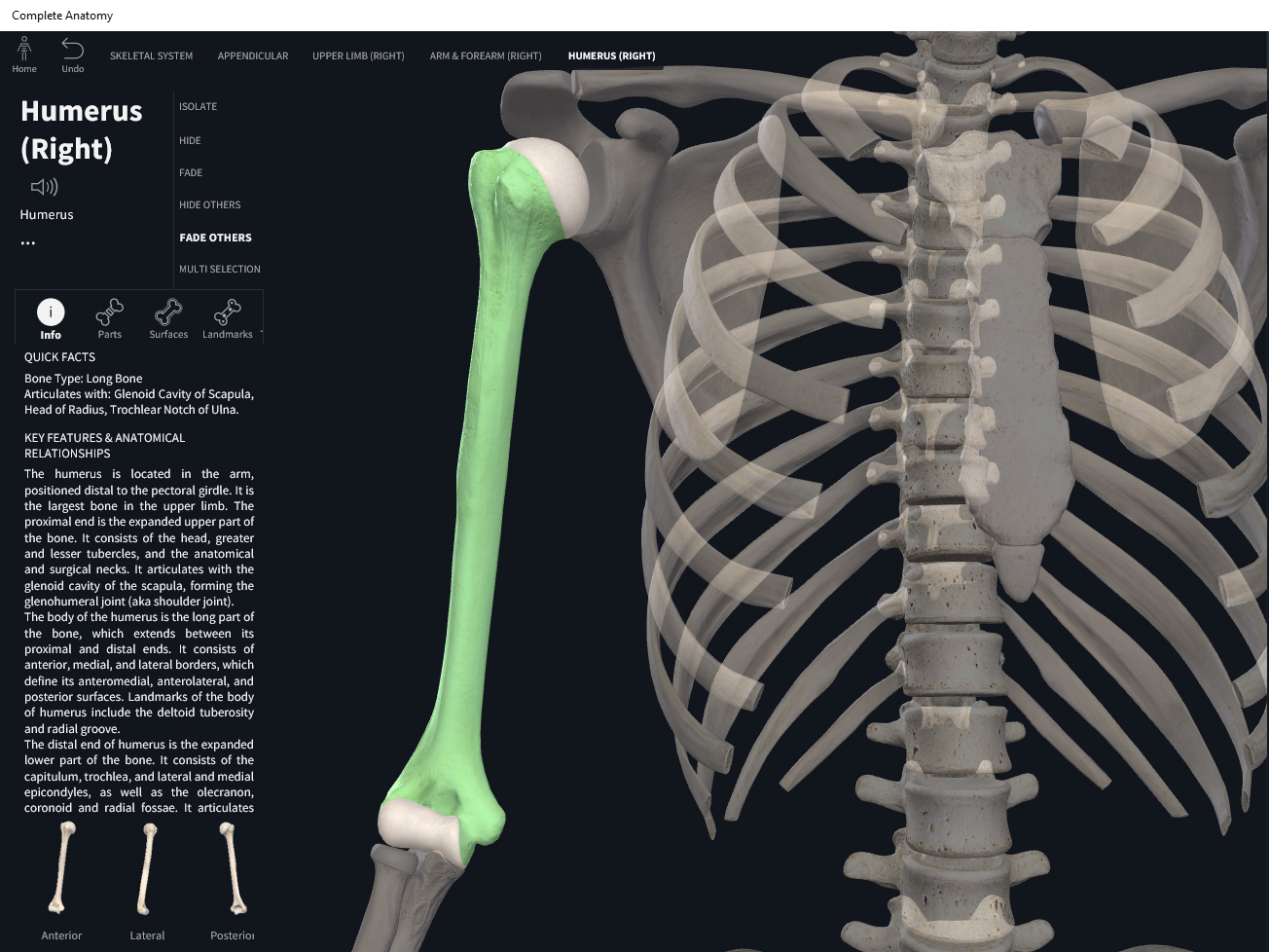
Bones: Humerus. – Anatomy & Physiology we have 9 Pictures about Bones: Humerus. – Anatomy & Physiology like Superior View of Medial Wall of Right Orbit and Frontal, Ethmoid, and, Superficial Musculature of the Posterior Neck | Neuroanatomy | The and also Superior View of Medial Wall of Right Orbit and Frontal, Ethmoid, and. Here it is:
Bones: Humerus. – Anatomy & Physiology
 integrativewellnessandmovement.com
humerus
AP View: Capitellum
 meds.queensu.ca
capitellum ap elbow ped radiograph radius queensu meds ts modules central
Image | Radiopaedia.org
 radiopaedia.org
pellegrini stieda lesion radiopaedia radiology medial femoral ligament collateral bones adjacent annotated lesions pinu
Medial Meniscus Cyst | Image | Radiopaedia.org
 radiopaedia.org
meniscus cyst medial knee pa radiopaedia meniscal tear leg standing synovial version
Superior View Of Medial Wall Of Right Orbit And Frontal, Ethmoid, And
 www.neurosurgicalatlas.com
ethmoid wall neuroanatomy
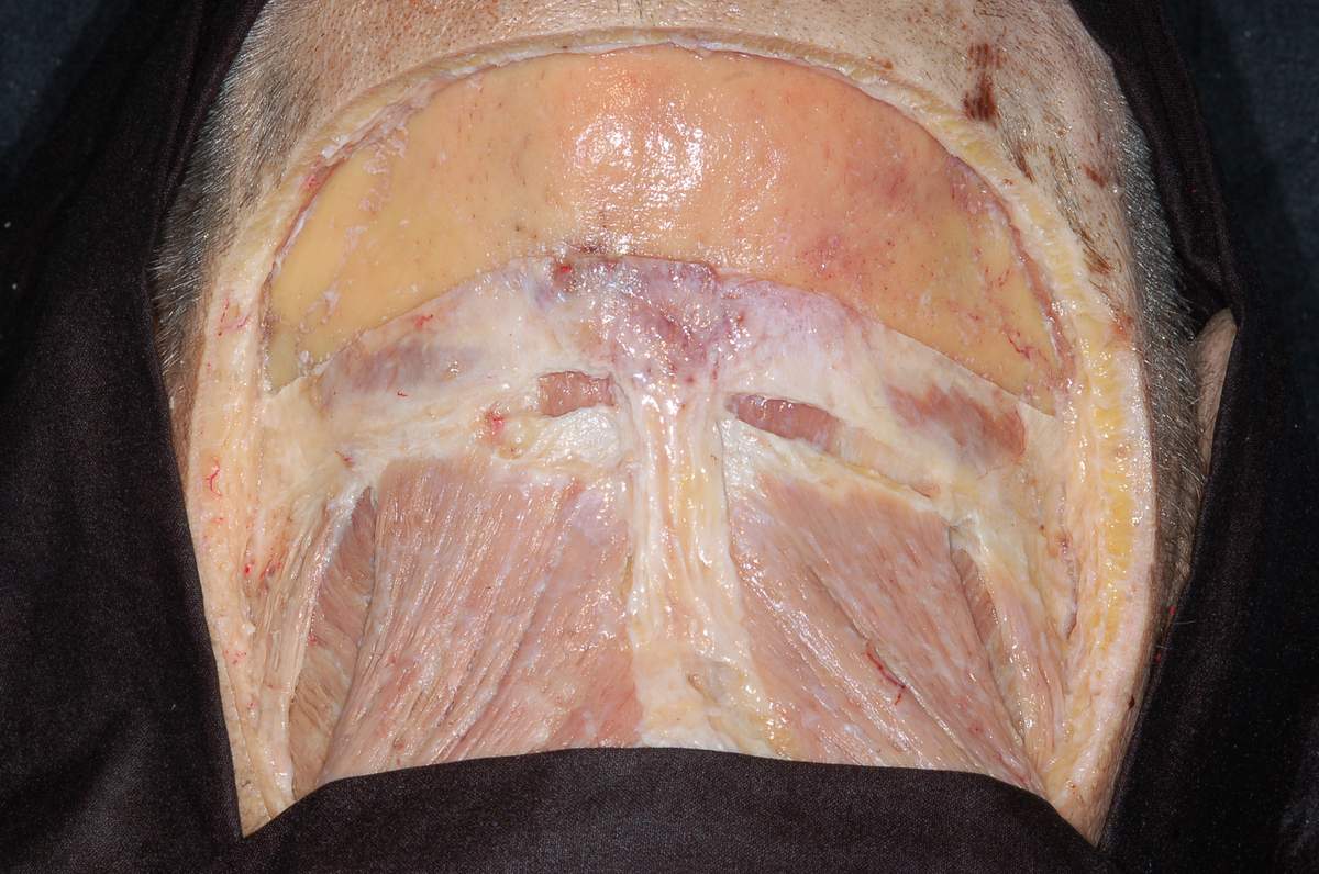
Superficial Musculature Of The Posterior Neck | Neuroanatomy | The
 www.neurosurgicalatlas.com
posterior superficial musculature neurosurgicalatlas surgical
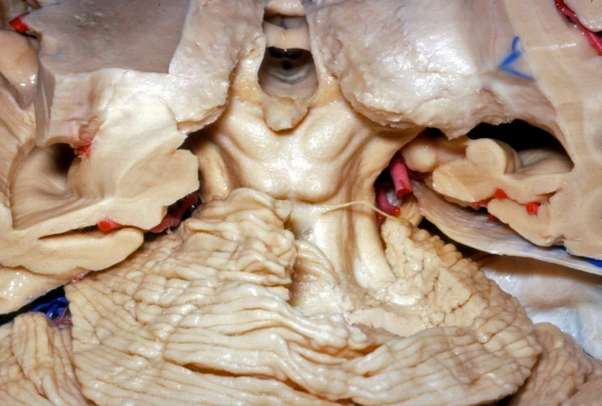
Posterior Perspective Of The Dissected Pineal Region | Neuroanatomy
 www.neurosurgicalatlas.com
pineal dissected neurosurgicalatlas correlation
Ischiofemoral Impingement Syndrome - Radsource
 radsource.us
ischiofemoral impingement space anatomy mri hip syndrome radsource ifi
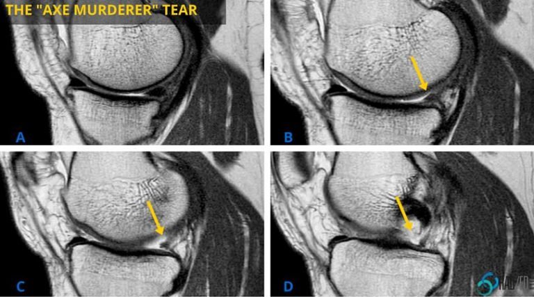
Meniscus Posterior Horn Part 1: An Axe Murderer Meets The Ghost
 radedasia.com
meniscus posterior horn tear root mri medial meniscal lateral ghost axe murderer sagittal intercondylar radedasia sag radiology meets fossa truncated
Ethmoid wall neuroanatomy. Pineal dissected neurosurgicalatlas correlation. Meniscus posterior horn tear root mri medial meniscal lateral ghost axe murderer sagittal intercondylar radedasia sag radiology meets fossa truncated