thoracic venous anatomy
Congenital Variations of TOS | Center for Thoracic Outlet Syndrome we have 9 Pictures about Congenital Variations of TOS | Center for Thoracic Outlet Syndrome like Thoracic duct: Anatomy, course and clinical significance | Kenhub, Blood Supply to the Spinal Cord Medical Illustration Medivisuals and also Cervical fascias: Superficial and deep fascial layers | Kenhub. Here you go:
Congenital Variations Of TOS | Center For Thoracic Outlet Syndrome
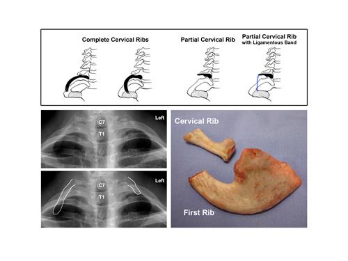 tos.wustl.edu
congenital tos variations cervical thoracic
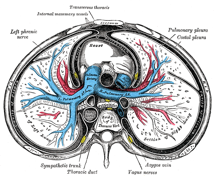
Cervical Fascias: Superficial And Deep Fascial Layers | Kenhub
sympathetic esophagus nervous system trunk anatomy cervical kenhub lateral plexus fascias brachial oesophagus autonomic pericardium drainage neurovascular parts lymphatic truncus
Superior Vena Cava - Wikidoc
 www.wikidoc.org
pulmonary artery anatomy lung arteries vessels embolism transverse vena section vein thoracic nerve superior thorax cava aorta relations bronchus azygos
Upper-extremity Deep Venous Thrombosis: A Review - The American Journal
 www.amjmed.com
venous ultrasound upper extremity anatomy veins brachiocephalic deep jugular thrombosis common figure axillary most
Anaesthesia UK : Anatomy Relevant To Epidural And Subarachnoid Blockade
 www.frca.co.uk
anatomy body spine spinal epidural human landmarks posterior scapula surface cord study subarachnoid ilium anaesthesia space markings blockade relevant spinous
Blood Supply To The Spinal Cord Medical Illustration Medivisuals
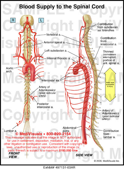 www.medivisuals1.com
spinal cord supply blood medical medivisuals1
Abdominal CT Anatomy | Radiology Key
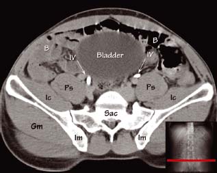 radiologykey.com
ct anatomy abdominal pelvis bladder radiology key radiologykey
Thoracic Duct: Anatomy, Course And Clinical Significance | Kenhub
thoracic lymph lymphatic kenhub tributaries münstermann irina
Duplex Assessment Of Deep Venous Thrombosis And Upper-limb Venous
 radiologykey.com
venous limb veins thrombosis radiology
Sympathetic esophagus nervous system trunk anatomy cervical kenhub lateral plexus fascias brachial oesophagus autonomic pericardium drainage neurovascular parts lymphatic truncus. Duplex assessment of deep venous thrombosis and upper-limb venous. Venous ultrasound upper extremity anatomy veins brachiocephalic deep jugular thrombosis common figure axillary most